Soon I begin a job radiographing yearling horses across NSW. I anticipate become more adept at reading radiographs, and at working with horses. Considering the nature of the work I feel I should probably revise radiology and radiography in order to make sure all the basics are there to begin with. So over the next few days I will be writing about x-rays, and reading about analysis of equine radiographs.
So how are X-rays made?
So how are X-rays made?
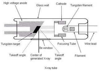 X-rays are electromagnetic radiation produced by electron interactions outside the nucleus. In order to produce this interaction an x-ray tube is used. The tube is made of glass and contains within it a wire filament cathode, and a tungsten anode (which may be stationary or rotating). An electric current is passed through the cathode filament causing it to heat up and release electrons into an electron cloud around the filament. The higher the mA, the greater the current passing through the filament, and therefore the more electrons in the cloud. The electron cloud remains stationary unless a voltage differential (kVp) exists between the cathode and the anode. Since electrons are negatively charged the positively charged anode attracts the electrons to it. The greater the potential difference (kVp) between the cathode and anode, the faster the velocity of the electrons. This means that the electrons strike the anode with greater energy and so the x-rays produced have higher energy. The greater the energy of an x-ray the greater its penetration of matter.
 The focal spot is the region on the anode target which is struck by electrons. It is the site of x-ray production. A smaller focal spot gives a better detailed radiograph than a large focal spot because of the penumbra effect.
Increasing mA increases the number of x-rays being produced. Increase the length of time the x-ray tube is energised also increases the number of x-rays produced. Therefore mAs is used to determine the total number of x-rays being produced. mAs = mA x time.
By changing mA, time, kVp, focal size, focal length the detail, penetration, and contrast of a radiograph can be altered.

Listening to "Hello its me" by Four Star Mary

No comments:
Post a Comment