I just read a facinating article by JH Creeper and NB Buller in the Australian Veterinary Journal (Nov 2006, 84 (11): 408-411) An outbreak of Streptococcus iniae in barramundi (Lates calcarifera) in fresh water culture cages. Having recently begun to learn about the application of terrestrial veterinary work into the aquaculture industry this is particularly interesting.
Fish are quite unique in that in terms of HPE interactions the environment has a very intimate relationship to the host. The nature of aquaculture systems means that strict attention must be paid to the environment, HACCAP principles should be applied in order to reduce the effect of environmental stressors on the fish, and hence decrease the risk of pathology.
The clinical signs shown by the fish studied in this outbreak were typical of those seen in septicaemia. Exopthalmia, reddening of the skin around fins and ventral abdomen, splenomegally, rapid respiration, and slow swimming near surface.
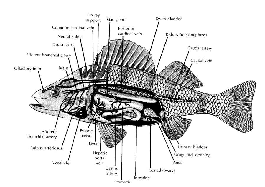
Necropsy is one of the most important disciplines of fish veterinary work, it should be done promptly on at least 3-5 moribund fish. As with all necropsy care must be taken to use sterile technique. Flash disinfect the fish with 70% ethanol solution. Using a heated spatula sear the ventral and lateral surfaces of the abdomen. Remove the entire lateral abdominal wall to expose the abdominal cavity using aseptic technique. Examine the abdominal contents visually, is it normal, what degree of autolysis exists, and any structures enlarged or abnormal?
Using rat tooth forceps grasp the caudal end of the swimbladder and reflect it rostally. This exposes the anterior/head kidney which is an ideal site for microbiological sampling. Using a sterile swab collect a sample from the anterior kidney and plate it out onto blood agar. The spleen may also be sampled if septicaemia is suspected.
Next sample the heart by using scissors to aseptically open the cardiac space. Assess appearance of heart and blood vessels. Then open the heart (preferably left ventricle as this is the largest) and sample the blood with a sterile swab. Plate this out. The heart should also be taken for histological analysis, so remove it and place it in formalin.
Sample the brain. Sear the Dorsal and dorsolateral surfaces of the cranium. Using aseptic technique dissect into the cranial cavity and reflect the bony dorsum from above the brain. Swab and plate out a sample from the brain. All cultures should be incubated for at
least 24 hours but up to 3 days is ideal.
Gill sampling is also very important. The gill should be analysed histologically. Take a wedge of gill using scissors and place in formalin.
In the discussion it is noted that environmental factors were important contributing factors to the Streptococcus iniae outbreak. Silt and mud entered the cages after rain introducing the bacterium, and then overstocking, hydrogen sulphide, high water temperatures (33 degrees Celsius), and low dissolved oxygen (less than or equal to 5mg/L) were contributing factors to the outbreak of disease.
This article was a really interesting read, and help me to revise my fish vet skills. Aquaculture is a booming industry and it is important that vet's get involved. It would be really interesting to get some fish work under my belt. I will have to investigate this area further.

No comments:
Post a Comment