Monday 20 November 2006

What happens to the x-ray when they pass through the patient?

There are five possible ways for the x-ray photon to interact with the matter of the patient (and they may do more than one type).


  1. Coherent Scatter - the photon hits the object and changes direction with no absorbtion of energy. This type of interact is detrimental to diagnostic radiography as they can strike the film and degrade image quality, or strike the radiographer exposing them to radiation

  2. Photoelectric Effect - the photon hits the patient ans is totally absorbed, and ejects and electron from an inner shell of a tissue atom, the space is then filled by another electron which causes a characteristic x-ray to be given off. The probability of this reaction occuring is in direct proportion to a tissues atomic number. It is this atomic number dependence on the photoelectric effect that creates the contrasts seen between the different opacities on an image.

  3. Compton Scattering - photon interacts with a peripheral shell electron, the electron is ejected and the phot is scattered at different angles. This is disadvantagous as it degrades the image and causes personell exposure.

  4. Pair production - no relevance to x-ray production

  5. Photodisintegration - no relevance to x-ray production


That's enough learning for today, I have other plans. This afternoon I am going to a BBQ with some friends of mine. Should be good, but no drinking because I have to drive (stupid zero limit!) We are suppose to bring our own meat but I don't know what I should bring? Its very hot so as long as I don't have to cook any thing is good I guess. Wish Ducky had a pool though.


Listening to The Killers "Sam's Town" but on the road I am thinking that Rocky Horror Picture Show or Pricilla Queen of the Desert Soundtracks might be the go. Feel like vamping!!


How are X-rays produced?


Soon I begin a job radiographing yearling horses across NSW. I anticipate become more adept at reading radiographs, and at working with horses. Considering the nature of the work I feel I should probably revise radiology and radiography in order to make sure all the basics are there to begin with. So over the next few days I will be writing about x-rays, and reading about analysis of equine radiographs.

So how are X-rays made?


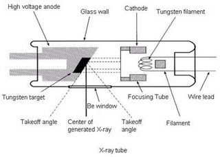
X-rays are electromagnetic radiation produced by electron interactions outside the nucleus. In order to produce this interaction an x-ray tube is used. The tube is made of glass and contains within it a wire filament cathode, and a tungsten anode (which may be stationary or rotating). An electric current is passed through the cathode filament causing it to heat up and release electrons into an electron cloud around the filament. The higher the mA, the greater the current passing through the filament, and therefore the more electrons in the cloud. The electron cloud remains stationary unless a voltage differential (kVp) exists between the cathode and the anode. Since electrons are negatively charged the positively charged anode attracts the electrons to it. The greater the potential difference (kVp) between the cathode and anode, the faster the velocity of the electrons. This means that the electrons strike the anode with greater energy and so the x-rays produced have higher energy. The greater the energy of an x-ray the greater its penetration of matter.


At the anode x-rays are produced by collisional and bremsstrahlung interactions. Collisional interactions occur when an oncoming electron ejects and orbital electron from the atom in the target with subsequent release of energy as an x-ray. In a bremsstrahlung interaction the oncoming electron slows as it bends around the nucleus because of the difference in charges, it releases energy(x-rays) as it is deflected.

The focal spot is the region on the anode target which is struck by electrons. It is the site of x-ray production. A smaller focal spot gives a better detailed radiograph than a large focal spot because of the penumbra effect.

Increasing mA increases the number of x-rays being produced. Increase the length of time the x-ray tube is energised also increases the number of x-rays produced. Therefore mAs is used to determine the total number of x-rays being produced. mAs = mA x time.

By changing mA, time, kVp, focal size, focal length the detail, penetration, and contrast of a radiograph can be altered.


Listening to "Hello its me" by Four Star Mary

Fish Vet??


I just read a facinating article by JH Creeper and NB Buller in the Australian Veterinary Journal (Nov 2006, 84 (11): 408-411) An outbreak of Streptococcus iniae in barramundi (Lates calcarifera) in fresh water culture cages. Having recently begun to learn about the application of terrestrial veterinary work into the aquaculture industry this is particularly interesting.


Fish are quite unique in that in terms of HPE interactions the environment has a very intimate relationship to the host. The nature of aquaculture systems means that strict attention must be paid to the environment, HACCAP principles should be applied in order to reduce the effect of environmental stressors on the fish, and hence decrease the risk of pathology.
The clinical signs shown by the fish studied in this outbreak were typical of those seen in septicaemia. Exopthalmia, reddening of the skin around fins and ventral abdomen, splenomegally, rapid respiration, and slow swimming near surface.
Necropsy is one of the most important disciplines of fish veterinary work, it should be done promptly on at least 3-5 moribund fish. As with all necropsy care must be taken to use sterile technique. Flash disinfect the fish with 70% ethanol solution. Using a heated spatula sear the ventral and lateral surfaces of the abdomen. Remove the entire lateral abdominal wall to expose the abdominal cavity using aseptic technique. Examine the abdominal contents visually, is it normal, what degree of autolysis exists, and any structures enlarged or abnormal?
Using rat tooth forceps grasp the caudal end of the swimbladder and reflect it rostally. This exposes the anterior/head kidney which is an ideal site for microbiological sampling. Using a sterile swab collect a sample from the anterior kidney and plate it out onto blood agar. The spleen may also be sampled if septicaemia is suspected.
Next sample the heart by using scissors to aseptically open the cardiac space. Assess appearance of heart and blood vessels. Then open the heart (preferably left ventricle as this is the largest) and sample the blood with a sterile swab. Plate this out. The heart should also be taken for histological analysis, so remove it and place it in formalin.
Sample the brain. Sear the Dorsal and dorsolateral surfaces of the cranium. Using aseptic technique dissect into the cranial cavity and reflect the bony dorsum from above the brain. Swab and plate out a sample from the brain. All cultures should be incubated for at
least 24 hours but up to 3 days is ideal.
Gill sampling is also very important. The gill should be analysed histologically. Take a wedge of gill using scissors and place in formalin.
In the discussion it is noted that environmental factors were important contributing factors to the Streptococcus iniae outbreak. Silt and mud entered the cages after rain introducing the bacterium, and then overstocking, hydrogen sulphide, high water temperatures (33 degrees Celsius), and low dissolved oxygen (less than or equal to 5mg/L) were contributing factors to the outbreak of disease.
This article was a really interesting read, and help me to revise my fish vet skills. Aquaculture is a booming industry and it is important that vet's get involved. It would be really interesting to get some fish work under my belt. I will have to investigate this area further.

Sunday 19 November 2006

Summary of corrective surgery for Patent Ductus Arteriosus

A patent ductus arteriosus is a congenital defect that occurs when the ductus arteriosus which connects the descending aorta and left pulmonary artery does not close. While it is an important structure in the foetus, after birth if it fails to close it can cause significant pathology. A continuous machinery murmur will be heard in patients with a PDA. The treatment of choice in small animals is ductus arteriosus ligation, which is considered to be curative.


Once anaesthetised prepare the dog for surgery by clipping up the thorax extending to mid abdomen, and to midway up chest wall. Clean the area using chlorhexidine, starting at the center and moving outwards. Follow up with iodine wash again moving from the centre to the outside.


Transport patient into surgery and position patient on right side in lateral recumbency for a left 4th intercostal thoractomy.


Scrub in using sterile procedures. Drape the patient using quarter drapes, secure the field drapes with backhaus towel clamps inorder to securely isolate the unprepared portion of the patient. Now place a large drape over the animal and surgical table to complete the sterile field.


Incise the skin, subcutaneous tissue and cutaneous trunci above 4th intercostal space running from the just ventral to the base of the vertebral bodies to just dorsal to the sternum. Using scissors incise through the latissimus dorsi muscle, and then transect the scalenus and pectoral muscles perpendicular to their fibres. Separate the serratus ventralis fibres above the intercostal space. Using scissors cut the external and then internal intercostal muscles near the costochondral junction.


You are now about to enter the thoracic cavity, so alert the anaesthetist to being ventilating the patient once the pleura is breached. Using blunt scissors penetrate the pleura. Now taking care to avoid the internal thoracic vessels extend the incision dorsally and ventrally to allow greater access to the thoracic cavity. Drape the edges of the incision with moistened labarotomy sponges and use a finochietto retractor to separate the ribs. The ribs will spread more easily cranial to the intercostal incision.


Now locate the heart in the thoracic cavity. Once this is done identify the left vagus nerve as it courses over the ductus arteriosus between the aorta and main pulmonary arteries. Isolate the vagus using sharp dissection as secure it away from the ductus arteriosus using a suture to gently retract it (watch for vagal induced bradycardia, or other vagus associated responses). Without entering the pericardial sac bluntly dissect around the PDA, right angled forceps are often useful for ispolating hte caudal and cranial aspects of the ductus. Pass the forceps from medial to the ductus in a caudocranial direction and grasp a piece of non-absorbable suture (heavy silk No. 1 or 0 is ideal). Slowly pull the suture beneath the ductus, repeat this again do not secure the suture. Now pass a second suture using the same technique. Now slowly tighten the suture closest to the aorta, and next the second suture closer to the pulmonary arteries. The ligation of the PDA is now complete.


Close the thoracic cavity by placing heavy non-absorbable suture around the ribs adjacent to the incision, but do not tie. Using a rib approximator, apose the ribs, now secure the pre-placed sutures. Remove the rib approximator. Suture first the serratus venetralis, then scalenus and pectoralis muscles using a continuous suture pattern in absorbable suture material. Now appose and continuously suture the edges of latissimus dorsi muscle. Remove any residual air from the thoracic cavity using an over-the-needle catheter. Close the subcutaneous tissue and skin using either a continuous or interrupted suture pattern.

Dreaming

I had a dream last night. I was in a 2nd hand book store standing in front of a shelf full of classics, looking for Jack London's "Call of the wild". Why did this cause me to begin this? Well I don't want my brain to turn into mush - and this was a subconscious call for help.

Now as I detox from too much caffeine, prepare for a early morning run with Rich, and attempt to get over a cold, I begin. Beware to all who read on it may get scary, it may get boring, it may get raunchy?! Where ever the mood takes me, you will go to.

Listening to Blondie "Call me"
Reading 'Paycheck' by Philip K. Dick (no wonder my mind is dying)